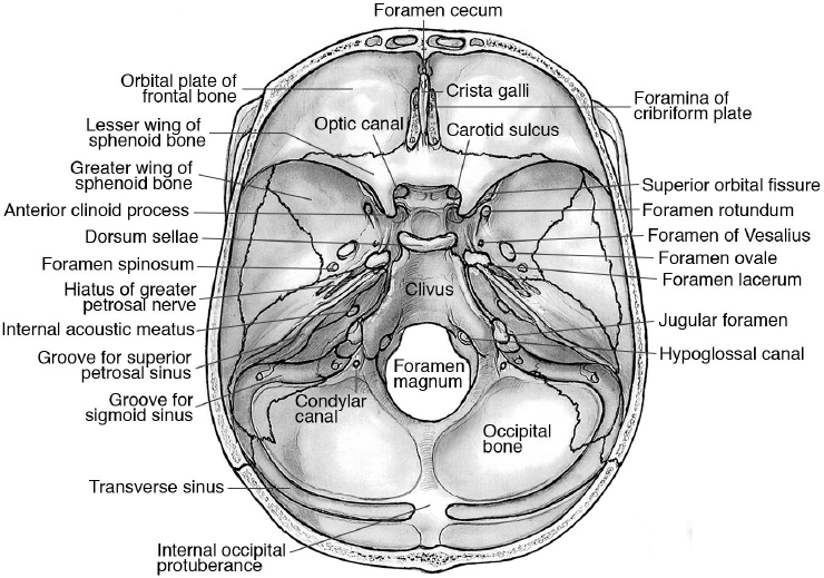
The parasellar structures include the sphenoidal sinus and adjoining central skull base the paired cavernous sinuses and adjoining venous sinuses the cavernous and supraclinoid segments of the internal carotid arteries the circle of willis the optic nerves and chiasm.
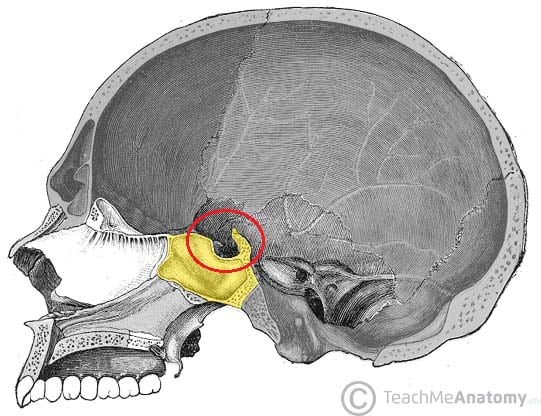
Floor view skull dorsunm salla.
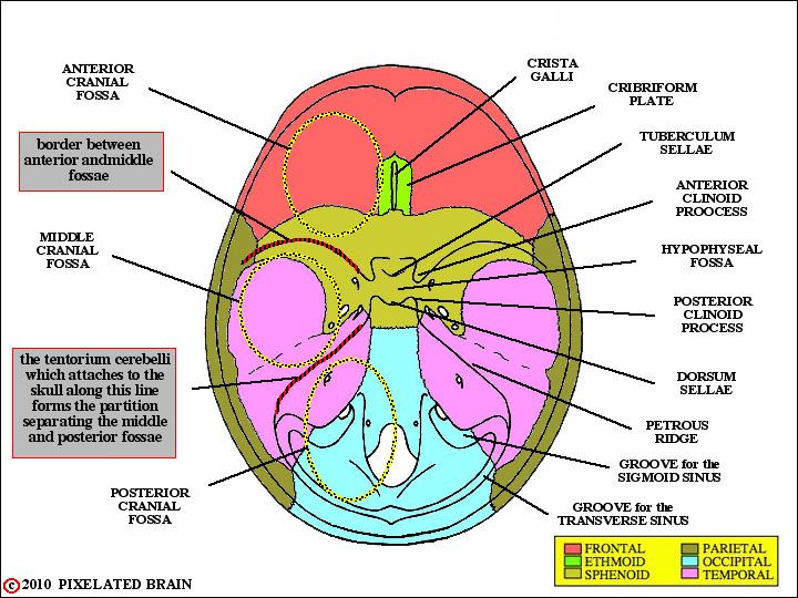
The cranium skull is the skeletal structure of the head that supports the face and protects the brain it is subdivided into the facial bones and the brain case or cranial vault figure 1 the facial bones underlie the facial structures form the nasal cavity enclose the eyeballs and support the teeth of the upper and lower jaws.
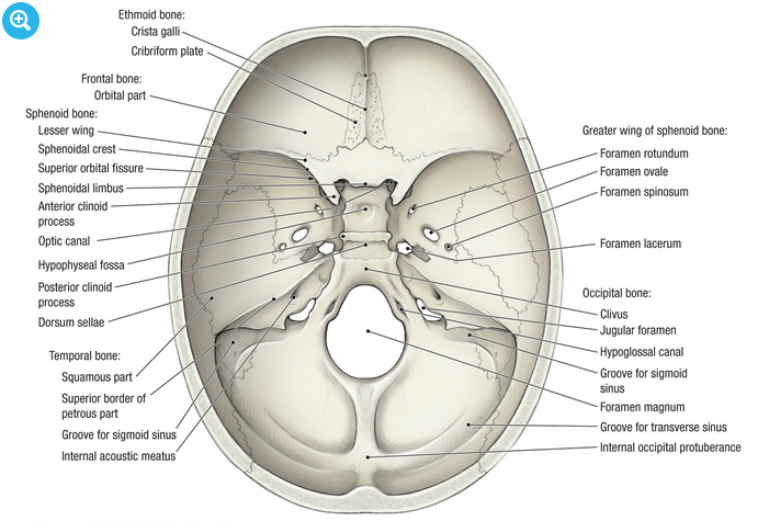
The dorsum sellae is part of the sphenoid bone in the skull together with the basilar part of the occipital bone it forms the clivus.
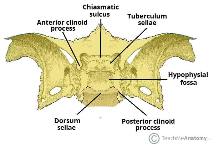
The dorsum sellae is the square shaped process of the sphenoid bone it ascends superiorly from the posterior part of the sphenoid body to form the posterior wall of the sella turcica.
Ballooned sella with an undercutting anterior clinoid process unequal downward displacement of the floor double floor appearance craniopharyngioma elongated sella with short curved dorsum is characteristic but more often indistinguishable from the pituitary lesions.
Functions of sella turcica.
The dura is left intact and the sellotomy extends from carotid artery c to carotid artery in a lateral dimension and from the tuberculum sella ts superiorly to the floor of the sella inferiorly.
Any anomaly or pathological condition in the pituitary gland can apparent from a distorted form of sella turcica to a disorder in the malfunctioning of the secretion of the hormones secreted by the pituitary which includes.
A normal sella turcica b oblique anterior wall c sella turcica bridge d double contour of floor e irregular dorsum sellae f pyramidal shape.
In the sphenoid bone the anterior boundary of the sella turcica is completed by two small eminences one on either side called the middle clinoid processes while the posterior boundary is formed by a square shaped plate of bone the dorsum sellae ending at.
The dorsum sellae forms the posterior wall of the sella turcica which houses the pituitary gland laterally it articulates with the petrous apex of the petrous part of the temporal bone.
Providing the proper place to hold and support the pituitary gland.
Location of sella turcica in skull.
The sella turcica latin for turkish seat is a saddle shaped depression in the body of the sphenoid bone of the human skull and of the skulls of other hominids including chimpanzees orangutans and gorillas it serves as a cephalometric landmark the pituitary gland or hypophysis is located within the most inferior aspect of the sella turcica the hypophyseal fossa.
Different morphological types of sella turcica according to axelson et al.
Empty sella is the designation for intrasellar herniation of the meningeal space which results in a neuroanatomical picture in which the pituitary gland appears to be thinned and flattened against the sellar floor the bulk of the sellar space being filled with csf kaufman et al.
B endoscopic view after removal of the posterior sphenoid base.