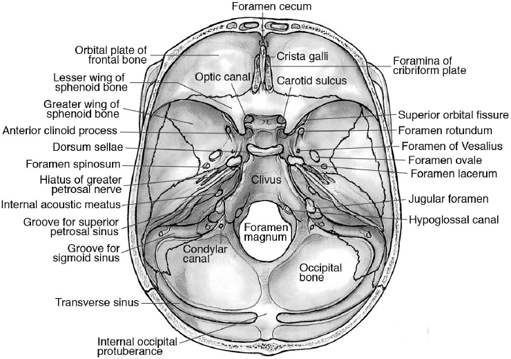
Floor View Skull Dorsum Sellae

Specialised Projections Of The Skull Radiology Key
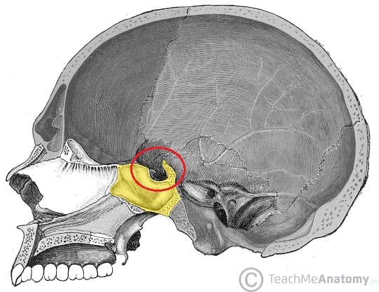
Middle Cranial Fossa Boundaries Contents Teachmeanatomy

Landmarks For Cephalometric Analysis S Sella Center Of Sella Download Scientific Diagram
Anatomy Of The Skull Base And Related Structures Elements Of Surgical Anatomy Neupsy Key

Skull X Ray Lateral View Note Enlargement Of Pituitary Fossa Loss Download Scientific Diagram
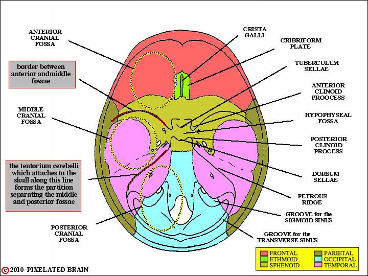
Pixelated Brain Module 1 Section 1 The Skull

In a properly positioned caldwell projection the ir is perpendicular to the orbitomeatal line oml and the x rays pass at an angle of 15 degrees from behind the head and exit at the nasion.
Floor view skull dorsum sellae.

The Axial Skeleton Flashcards Quizlet
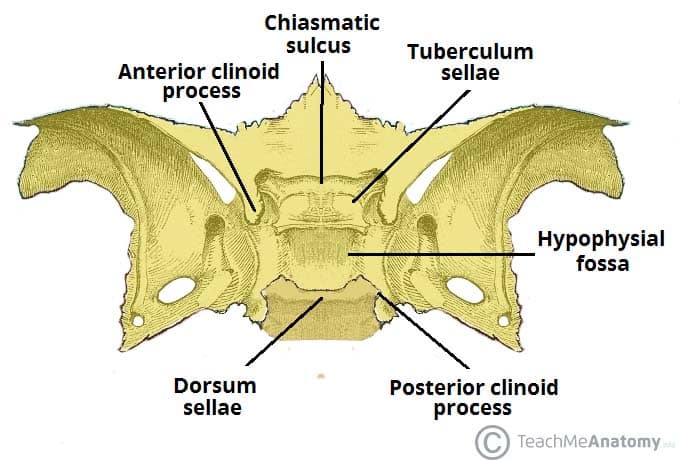
Sphenoid Bone Location Structure Function Teachmeanatomy
Skull Foramina Fissures And Contents Kenhub
Sphenoid Bone
Source : pinterest.com
